topic 2: Muscles and Energy
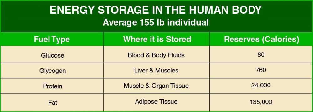
Muscles use the stored chemical energy from food we eat and convert that to heat and energy of motion (kinetic energy). Energy is required to enable growth and repair of tissue, to maintain body temperature and to fuel physical activity. Energy comes from foods rich in carbohydrate, protein and fat.
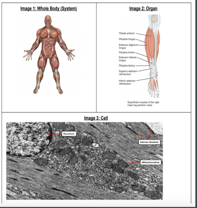
Identify the levels of organization of muscles
Identify: Find an answer from a given number of possibilities
Identify: Find an answer from a given number of possibilities
- Molecular level: This level of organization involves the smallest functional units of muscle tissue, which are individual molecules such as actin and myosin.
- Cellular level: At this level of organization, muscle tissue is composed of individual muscle cells or muscle fibers. These cells are specialized for contraction and are characterized by the presence of myofibrils, which are made up of sarcomeres.
- Tissue level: This level of organization involves the grouping of muscle cells into larger structures called muscle tissue. Muscle tissue is composed of individual muscle fibers that are organized into fascicles.
- Organ level: At this level of organization, muscle tissue is further organized into functional units called muscles or muscle organs. A muscle organ is composed of muscle tissue, blood vessels, and nerves, and is responsible for producing movement.
- System level: At the highest level of organization, muscles work together with other organs and tissues to form functional systems such as the muscular, skeletal, and nervous systems. These systems work together to produce coordinated movement and maintain bodily functions
Identify the different types of muscle tissue, including skeletal, smooth, and cardiac muscle, and understanding their unique characteristics and functions.
Identify: Find an answer from a given number of possibilities
Identify: Find an answer from a given number of possibilities
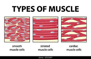
Skeletal muscle: Skeletal muscle is attached to bones and is responsible for voluntary movements of the body. It is characterized by its striated appearance, which is caused by the alignment of sarcomeres within the muscle fibers. Skeletal muscle is under conscious control and can be trained to increase strength and endurance.
Smooth muscle: Smooth muscle is found in the walls of organs and structures such as blood vessels, the digestive tract, and the uterus. It is responsible for involuntary movements such as peristalsis and vasoconstriction. Smooth muscle is not striated and has a more spindle-like shape than skeletal muscle.
Cardiac muscle: Cardiac muscle is found in the walls of the heart and is responsible for pumping blood throughout the body. It is characterized by its branching structure and intercalated discs, which allow for rapid conduction of electrical signals within the heart. Like skeletal muscle, cardiac muscle is striated, but it is involuntary and under the control of the autonomic nervous system.
Explain muscle action in terms of inputs and outputs
Explain: Give a detailed account of causes, reasons or mechanisms
Explain: Give a detailed account of causes, reasons or mechanisms
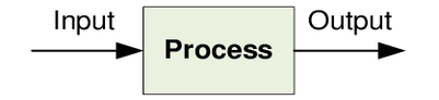
Inputs:
- Glucose: Glucose is a sugar that is the primary source of energy for muscle contraction. It is obtained from the bloodstream or stored in the muscle as glycogen.
- Oxygen: Oxygen is required for aerobic respiration, which produces ATP, the energy source for muscle contraction. Oxygen is obtained from the air through breathing and is transported to the muscle tissue via the bloodstream.
- ATP: ATP is the energy currency of the cell and is produced by aerobic and anaerobic respiration. ATP is used to fuel muscle contractions and other cellular processes.
- Water: Water is necessary for the hydration and functioning of muscle tissue. It is obtained from the bloodstream and through drinking fluids.
- CO2: CO2 is a waste product of cellular respiration and is produced by the muscles during contraction. It is transported to the lungs via the bloodstream and is exhaled.
- Movement or force: Movement or force is the output of muscle action. When a muscle contracts, it exerts a force on the bones or joints it is attached to, causing movement.
- ATP: ATP is produced by the muscle during cellular respiration and is used to fuel muscle contractions and other cellular processes.
- CO2: CO2 is a waste product of cellular respiration and is produced by the muscles during contraction. It is transported to the lungs via the bloodstream and is exhaled.
- Water: Water is produced by the muscles during cellular respiration and is necessary for the hydration and functioning of muscle tissue.
- Lactate: Lactate is produced by anaerobic respiration, which occurs when there is not enough oxygen available for aerobic respiration. Lactic acid can build up in the muscles, causing fatigue and soreness.
Analyze the relationship between exercise and cellular respiration, including how exercise affects the body's oxygen and glucose needs.
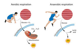
Cellular respiration is the process by which cells generate energy from glucose and other nutrients in the presence of oxygen. During cellular respiration, oxygen is used to break down glucose molecules and release energy in the form of ATP, which can then be used by cells to perform various functions, including muscle contraction.
Exercise involves physical activity that increases the body's demand for energy. This demand is met by an increased rate of cellular respiration, which allows cells to generate more ATP to meet the energy needs of the body. During exercise, the body's oxygen and glucose needs increase in order to fuel the increased rate of cellular respiration.
In the absence of sufficient oxygen, cells can undergo anaerobic respiration, which generates ATP in the absence of oxygen but produces lactic acid as a byproduct. This can lead to muscle fatigue and soreness.
Regular exercise can also lead to adaptations in the body that improve cellular respiration, such as an increase in the number and efficiency of mitochondria (the organelles responsible for cellular respiration) in muscle cells. This can lead to improved endurance and overall physical fitness.
Compare and contrast the different types of exercise, including aerobic, anaerobic, and resistance training, in terms of their energy requirements and impact on the body.
Aerobic exercise, anaerobic exercise, and resistance training are all different types of exercise that have different energy requirements and impacts on the body.
In terms of energy requirements, aerobic exercise requires a steady supply of oxygen and glucose to produce energy, while anaerobic exercise relies on stored energy sources and produces energy quickly without oxygen. Resistance training primarily uses anaerobic respiration and requires energy to be stored in the muscles themselves, in the form of glycogen.
In terms of impact on the body, aerobic exercise is effective for improving cardiovascular health, endurance, and weight loss. Anaerobic exercise is effective for building strength, power, and muscle mass, and can also improve cardiovascular health. Resistance training is effective for building muscle strength, improving bone density, and reducing the risk of injury.
- Aerobic exercise, also known as cardio exercise, is any exercise that involves sustained, rhythmic movements that increase heart rate and breathing, such as running, cycling, or swimming. This type of exercise primarily uses aerobic respiration, which requires oxygen and glucose to produce energy. Aerobic exercise is effective for improving cardiovascular health, endurance, and weight loss.
- Anaerobic exercise, on the other hand, involves short, intense bursts of activity that do not require oxygen and rely primarily on anaerobic respiration, which produces energy quickly but is less efficient than aerobic respiration. Examples of anaerobic exercise include sprinting, high-intensity interval training (HIIT), and weightlifting. This type of exercise is effective for building strength, power, and muscle mass.
- Resistance training, also known as strength training, involves the use of weights, resistance bands, or bodyweight to build strength and muscle mass. Resistance training primarily uses anaerobic respiration and involves working against a force or weight, such as lifting weights or doing push-ups. This type of exercise is effective for building muscle strength, improving bone density, and reducing the risk of injury.
In terms of energy requirements, aerobic exercise requires a steady supply of oxygen and glucose to produce energy, while anaerobic exercise relies on stored energy sources and produces energy quickly without oxygen. Resistance training primarily uses anaerobic respiration and requires energy to be stored in the muscles themselves, in the form of glycogen.
In terms of impact on the body, aerobic exercise is effective for improving cardiovascular health, endurance, and weight loss. Anaerobic exercise is effective for building strength, power, and muscle mass, and can also improve cardiovascular health. Resistance training is effective for building muscle strength, improving bone density, and reducing the risk of injury.
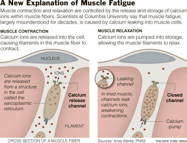
Describe the causes of muscle fatigue, including factors such as energy depletion, accumulation of metabolic byproducts, and damage to muscle fibers.
Describe: Give a detailed account
Describe: Give a detailed account
- Energy depletion: Muscle cells rely on the molecule ATP (adenosine triphosphate) to fuel muscle contractions. During prolonged or intense muscle activity, the demand for ATP can exceed the rate of production, leading to energy depletion. When ATP levels drop, the muscle fibers are unable to contract efficiently, leading to muscle fatigue.
- Accumulation of metabolic byproducts: During muscle activity, the cells produce metabolic byproducts such as lactic acid, which can accumulate in the muscle tissue. These byproducts can disrupt the normal cellular processes within the muscle fibers, leading to fatigue. In addition, the accumulation of hydrogen ions in the muscle tissue can decrease the pH of the muscle, further impairing muscle function.
- Damage to muscle fibers: Prolonged or intense muscle activity can cause damage to the muscle fibers, including micro-tears in the muscle tissue. This damage can trigger an inflammatory response, leading to swelling and pain in the affected muscles. This can contribute to muscle fatigue.
Other factors that can contribute to muscle fatigue include dehydration, electrolyte imbalances, and inadequate nutrition. Inadequate sleep and stress can also contribute to muscle fatigue.
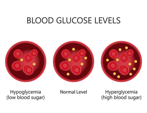
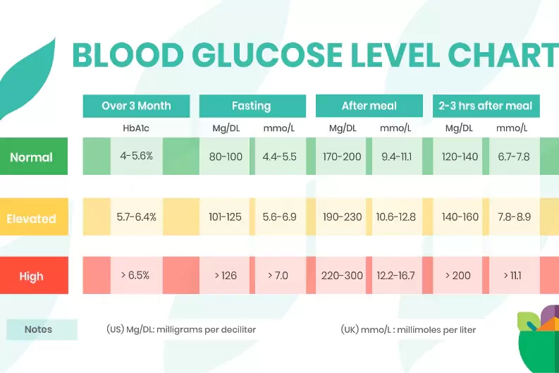
State that homeostasis is the maintenance of a constant internal environment and that blood glucose is an example of homeostasis
State: Give a specific name, value or other brief answer without explanation or calculation
State: Give a specific name, value or other brief answer without explanation or calculation
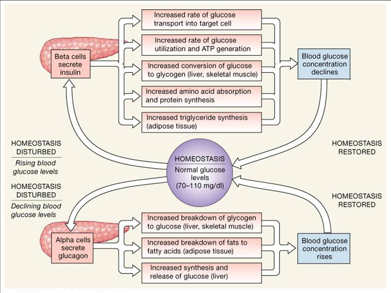
Homeostasis is the process of maintaining a relatively constant internal environment in the body despite changes in the external environment. This is achieved through a variety of regulatory mechanisms that help to balance and maintain the body's internal conditions.
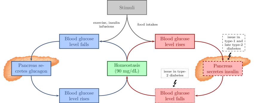
Blood glucose regulation is an example of homeostasis in the body. The body needs to maintain a relatively constant level of glucose in the bloodstream in order to provide energy to cells and tissues. When blood glucose levels rise, such as after a meal, the pancreas releases insulin to signal cells to take up glucose and store it for later use. When blood glucose levels drop, such as during fasting or exercise, the pancreas releases glucagon to signal the liver to convert stored glycogen into glucose and release it into the bloodstream.
By regulating blood glucose levels, the body is able to maintain a relatively constant internal environment that is necessary for the proper functioning of cells and tissues. Homeostasis is a critical component of overall health and well-being, and is involved in many other physiological processes in the body.
Explain how the body avoids having too little or too much glucose in the blood
Explain: Give a detailed account of causes, reasons or mechanisms
Explain: Give a detailed account of causes, reasons or mechanisms
The body has several mechanisms in place to ensure that the level of glucose in the blood is maintained within a narrow range, neither too little nor too much. This is important because glucose is the primary source of energy for the body's cells, and both low and high blood glucose levels can have negative effects on health.
The body avoids having too little glucose in the blood through a process called glycogenolysis, which is the breakdown of stored glycogen (a form of glucose storage in the liver and muscles) into glucose. This provides a quick source of glucose when blood glucose levels are low. The pancreas also releases the hormone glucagon, which signals the liver to convert stored glycogen into glucose and release it into the bloodstream. This raises blood glucose levels and provides energy to the cells.
On the other hand, the body avoids having too much glucose in the blood through a process called glycogenesis, which is the storage of glucose as glycogen in the liver and muscles. The hormone insulin, which is released by the pancreas in response to high blood glucose levels, signals cells to take up glucose from the bloodstream and convert it to glycogen for storage. This lowers blood glucose levels and prevents excessive glucose from damaging cells and tissues.
In addition to these mechanisms, the body also has a feedback system that involves the hypothalamus, the pituitary gland, and the adrenal glands. This system helps to regulate blood glucose levels by releasing hormones such as cortisol and epinephrine in response to changes in blood glucose levels. These hormones help to raise blood glucose levels when they are too low, and lower them when they are too high.
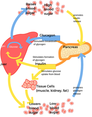
Draw a feedback mechanism for blood glucose regulation
Describe the function of the pancreas in regulating blood glucose
Describe: Give a detailed account
Describe: Give a detailed account
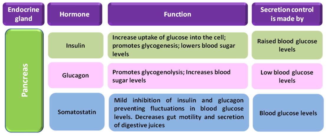
The pancreas is an organ located behind the stomach that plays an important role in regulating blood glucose levels in the body. It performs two main functions related to glucose metabolism: producing hormones that control blood glucose levels and producing enzymes that help digest food.
The pancreas contains clusters of cells called islets of Langerhans, which are responsible for producing the hormones insulin and glucagon. These hormones are released into the bloodstream in response to changes in blood glucose levels and work together to maintain blood glucose homeostasis.
Insulin is released by the pancreas in response to high blood glucose levels, such as after eating a meal. Insulin signals cells in the body to take up glucose from the bloodstream and convert it into energy or store it as glycogen for future use. This helps to lower blood glucose levels and prevent hyperglycemia (high blood glucose levels), which can lead to damage to organs and tissues in the body.
Glucagon, on the other hand, is released by the pancreas in response to low blood glucose levels, such as during fasting or exercise. Glucagon signals the liver to convert stored glycogen into glucose and release it into the bloodstream, which helps to raise blood glucose levels and prevent hypoglycemia (low blood glucose levels), which can lead to fatigue, dizziness, and other symptoms.
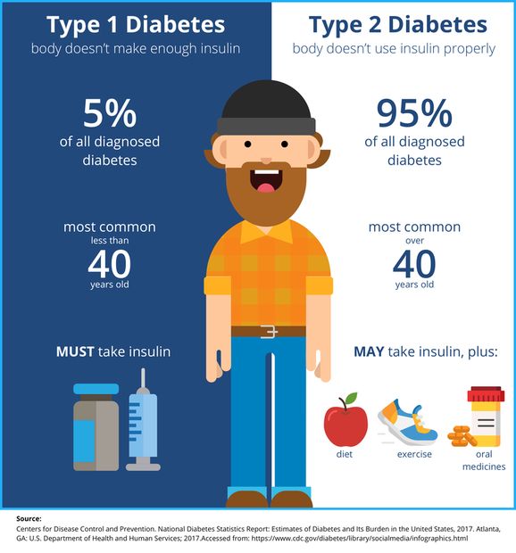
Outline the two different types of diabetes
Outline: Give a brief account
Outline: Give a brief account
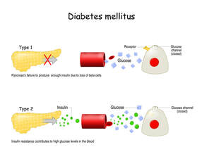
Diabetes occurs when the body is no longer able to control the level of glucose in the blood.
In general, people with diabetes either have a total lack of insulin (type 1 diabetes) or they have too little insulin or cannot use insulin effectively (type 2 diabetes).
In type 1 diabetes, the body's immune system destroys the cells that release insulin, eventually eliminating insulin production from the body. Without insulin, cells cannot absorb sugar (glucose), which they need to produce energy.
Type 2 diabetes can develop at any age. It most commonly becomes apparent during adulthood. But type 2 diabetes in children is rising. Type 2 diabetes accounts for the vast majority of people who have diabetes—90 to 95 out of 100 people. In type 2 diabetes, the body isn't able to use insulin the right way. This is called insulin resistance. As type 2 diabetes gets worse, the pancreas may make less and less insulin. This is called insulin deficiency.
Analyze the effects of different foods and macronutrients on blood glucose levels, including carbohydrates, proteins, and fats.
Analyze: Interpret data to reach conclusions
Analyze: Interpret data to reach conclusions
Different foods and macronutrients can have different effects on blood glucose levels, which can have important implications for overall health and disease risk.
- Carbohydrates are the primary macronutrient that can significantly affect blood glucose levels. When consumed, carbohydrates are broken down into glucose, which is then absorbed into the bloodstream and used by cells for energy. Simple carbohydrates, such as those found in sugary drinks and processed foods, are quickly absorbed and can cause a rapid increase in blood glucose levels. Complex carbohydrates, such as those found in whole grains and vegetables, are digested more slowly and cause a more gradual increase in blood glucose levels.
- Proteins and fats have less of an impact on blood glucose levels than carbohydrates. However, consuming large amounts of protein or fat without any carbohydrates can cause a slight increase in blood glucose levels due to a process called gluconeogenesis, in which the liver converts amino acids or fatty acids into glucose.
- Fiber is another important component of food that can affect blood glucose levels. Soluble fiber, which is found in foods such as oats, beans, and some fruits, can slow down the absorption of glucose and help to stabilize blood glucose levels. Insoluble fiber, which is found in foods such as whole grains and vegetables, can also slow down the absorption of glucose and promote overall digestive health.
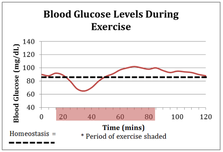
Evaluate changes in blood glucose due to eating, fasting, and exercising
Evaluate:Assess the implications and limitations
Evaluate:Assess the implications and limitations
- Eating: After eating, blood glucose levels typically rise as the carbohydrates in the food are digested and absorbed into the bloodstream. This triggers the pancreas to release insulin, which signals cells to take up glucose from the bloodstream and convert it to glycogen for storage. The rise in blood glucose levels after a meal is known as postprandial glucose, and it typically peaks around 1-2 hours after eating before gradually returning to baseline levels. The amount and type of food consumed can influence the magnitude and duration of the postprandial glucose response. Eating a meal high in carbohydrates and low in fiber, protein, and fat can lead to a more rapid and sustained rise in blood glucose levels.
- Fasting: Fasting, which involves abstaining from food and drink for a period of time, can lead to a decrease in blood glucose levels. This occurs because the body relies on stored glycogen in the liver and muscles to maintain blood glucose levels when food is not available. However, if fasting continues for an extended period of time, the liver will begin to convert stored glycogen into glucose and release it into the bloodstream to prevent hypoglycemia (low blood glucose). Prolonged fasting or starvation can lead to significant changes in blood glucose levels and can be detrimental to overall health.
- Exercise: Exercise can have both short-term and long-term effects on blood glucose levels. During exercise, the muscles require more energy and therefore take up more glucose from the bloodstream. This can lead to a temporary decrease in blood glucose levels. However, as exercise continues, the liver will begin to convert stored glycogen into glucose and release it into the bloodstream to maintain blood glucose levels. Over time, regular exercise can also increase insulin sensitivity, which can improve the body's ability to regulate blood glucose levels.
Discuss how fat is used as a storage molecule
Discuss: Give an account including, where possible, a range of arguments for and against the relative importance of various factors, or comparisons of alternative hypotheses
Discuss: Give an account including, where possible, a range of arguments for and against the relative importance of various factors, or comparisons of alternative hypotheses
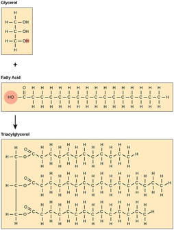
Fat is a type of nutrient that is used by the body as an energy source and a storage molecule. When the body consumes more calories than it needs for immediate energy, the excess calories are stored as fat for future use. Fat is stored in adipose tissue, which is found throughout the body, including under the skin and around organs.
Fat is an efficient storage molecule because it contains more energy per unit of weight than other storage molecules, such as carbohydrates. Fat molecules are made up of fatty acids and glycerol, which can be broken down and used for energy when needed. When the body needs energy, enzymes break down the fat molecules into fatty acids and glycerol, which are then transported to the cells and converted to ATP, the body's energy currency.
Fat is an efficient storage molecule because it contains more energy per unit of weight than other storage molecules, such as carbohydrates. Fat molecules are made up of fatty acids and glycerol, which can be broken down and used for energy when needed. When the body needs energy, enzymes break down the fat molecules into fatty acids and glycerol, which are then transported to the cells and converted to ATP, the body's energy currency.
Describe the circulatory system as a system of tubes with a pump and valves to ensure one way flow of blood
throughout the body
Describe: Give a detailed account
throughout the body
Describe: Give a detailed account
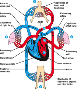 image www.haikudeck.com
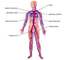
The circulatory system is the system of organs that circulates blood and lymph through the body, consisting of the heart, blood vessels, blood, lymph, and the lymphatic vessels and glands. The structure of the circulatory system is a blood vessels that transports nutrients and oxygen throughout the body.
Blood travels in a circular path through the circulatory system. The heart pumps blood to all parts of the body, and within about a minute, that blood returns to the heart to be pumped out once again. All cells of the body get their resources from the circulatory system, either directly or indirectly, depending on their proximity to blood vessels. Within the structure of the circulatory system, the heart is its center.
Blood travels in a circular path through the circulatory system. The heart pumps blood to all parts of the body, and within about a minute, that blood returns to the heart to be pumped out once again. All cells of the body get their resources from the circulatory system, either directly or indirectly, depending on their proximity to blood vessels. Within the structure of the circulatory system, the heart is its center.
Describe the structure of the heart including the muscular wall and septum, chambers, valves and associated blood vessels.
Describe: Give a detailed account
Describe: Give a detailed account
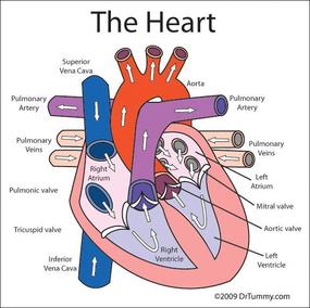
The heart pumps blood to all parts of the body, and within about a minute, that blood returns to the heart to be pumped out once again. All cells of the body get their resources from the circulatory system, either directly or indirectly, depending on their proximity to blood vessels.Within the structure of the circulatory system, the heart is its center.
he heart is made up of four different blood-filled areas, and each of these areas is called a chamber. There are two chambers on each side of the heart. One chamber is on the top and one chamber is on the bottom. The two chambers on top are called the atria. If you're talking only about one, call it an atrium. The atria are the chambers that fill with the blood returning to the heart from the body and lungs. The heart has a left atrium and a right atrium. heart diagram animated.
The two chambers on the bottom are called the ventricles The heart has a left ventricle and a right ventricle. Their job is to squirt out the blood to the body and lungs. Running down the middle of the heart is a thick wall of muscle called the septum. The septum's job is to separate the left side and the right side of the heart.
The atria and ventricles work as a team — the atria fill with blood, then dump it into the ventricles. The ventricles then squeeze, pumping blood out of the heart. While the ventricles are squeezing, the atria refill and get ready for the next contraction.
Describe the structure and functions of arteries, veins and capillaries
Describe: Give a detailed account
Describe: Give a detailed account
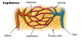
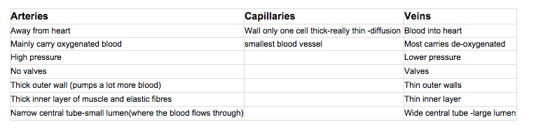
There are three kinds of blood vessels: arteries, veins, and capillaries. Each of these plays a very specific role in the circulation process.
Arteries carry oxygenated blood away from the heart. They’re tough on the outside but they contain a smooth interior layer of epithelial cells that allows blood to flow easily. Arteries also contain a strong, muscular middle layer that helps pump blood through the body.
Capillaries connect the arteries to veins. The arteries deliver the oxygen-rich blood to the capillaries, where the actual exchange of oxygen and carbon dioxide occurs. The capillaries then deliver the waste-rich blood to the veins for transport back to the lungs and heart.
Veins carry the blood back to the heart. They’re similar to arteries but not as strong or as thick. Unlike arteries, veins contain valves that ensure blood flows in only one direction. (Arteries don’t require valves because pressure from the heart is so strong that blood is only able to flow in one direction.) Valves also help blood travel back to the heart against the force of gravity.
Describe the transfer of materials between capillaries and tissue (diffusion and osmosis)
Describe: Give a detailed account
Describe: Give a detailed account
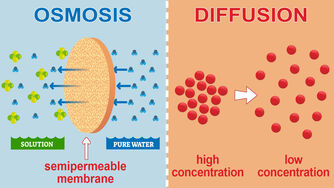
The transfer of materials between capillaries and tissue occurs primarily through the processes of diffusion and osmosis. Capillaries are the smallest blood vessels in the body and are responsible for delivering oxygen and nutrients to the tissues, as well as removing waste products.
Diffusion is the process by which molecules move from an area of higher concentration to an area of lower concentration. Oxygen and nutrients in the capillaries diffuse into the tissues, where their concentration is lower, while waste products such as carbon dioxide and urea diffuse out of the tissues into the capillaries, where their concentration is higher. The rate of diffusion is affected by factors such as the concentration gradient, the size and shape of the molecules, and the temperature.
Osmosis is a special type of diffusion that involves the movement of water molecules across a semi-permeable membrane. In the capillaries, water moves out of the bloodstream and into the tissues by osmosis, driven by the concentration of dissolved substances such as salts and sugars in the tissue fluid. This movement of water helps to maintain the balance of fluids between the capillaries and tissues.
Other factors that can affect the transfer of materials between capillaries and tissue include blood pressure, which can force fluids and molecules through the capillary walls, and the presence of specific transport proteins that help to move certain molecules across the membrane.
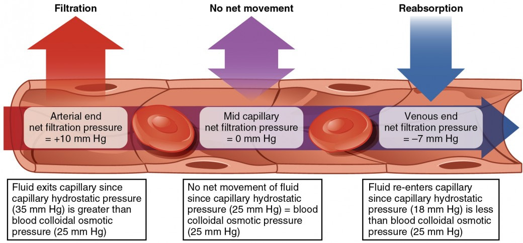
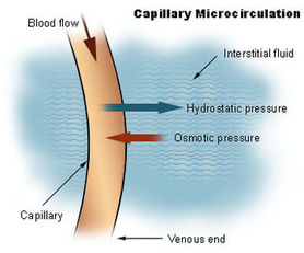 image from en.wikipedia.org
The blood capillaries are where the important functions of the circulation take place: the exchange of material between circulation and cells. Capillaries are the smallest of the body's blood vessels. They are only one cell thick, and they are the sites of the transfer of oxygen and other nutrients from the bloodstream to other tissues in the body; they also collect carbon dioxide waste materials and fluids for return to the veins. They connect the tiny muscular branches of arteries, called arterioles, with tiny veins (called venules). Ultimately, the capillary is the site of internal or cellular respiration and is responsible for the utilization of oxygen by the tissue and the transporting of carbon dioxide as waste to the veins for elimination by the lungs
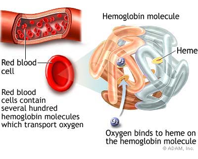
State the functions of red blood cells
State: Give a specific name, value or other brief answer without explanation or calculation
State: Give a specific name, value or other brief answer without explanation or calculation
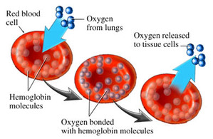 image from www.siliconvalleyfit.com
Red cells contain a special protein called hemoglobin, which helps carry oxygen from the lungs to the rest of the body and then returns carbon dioxide from the body to the lungs so it can be exhaled. Blood appears red because of the large number of red blood cells, which get their color from the hemoglobin. The percentage of whole blood volume that is made up of red blood cells is called the hematocrit and is a common measure of red blood cell levels.
Identify plasma as the liquid part of the blood.
Identify: Find an answer from a given number of possibilities
Identify: Find an answer from a given number of possibilities
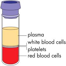 image from funwithmedicine.tumblr.com
Plasma is the clear, straw-colored liquid portion of blood that remains after red blood cells, white blood cells, platelets and other cellular components are removed. It is the single largest component of human blood, comprising about 55 percent, and contains water, carbon dioxide, salts, enzymes, antibodies and other proteins.
Composed of 90% water, plasma is a transporting medium for cells and a variety of substances vital to the human body.
Plasma carries out a variety of functions in the body, including clotting blood, fighting diseases and other critical functions.
Composed of 90% water, plasma is a transporting medium for cells and a variety of substances vital to the human body.
Plasma carries out a variety of functions in the body, including clotting blood, fighting diseases and other critical functions.
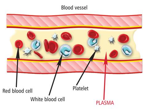
image from biotestplasma.com
Key Terms
|
carbon dioxide
pulse rate bpm blood pressure osmosis glycogen |
oxygen
carbon dioxide muscle heart blood glucagon |
artery
vein capillaries pancreas Type 1 Diabetes |
vessels
plasma RBC liver Type 2 Diabetes |
circulation
diffusion absorption glucose lipids |
Class Assignments
Useful Links
Videos
Videos
Biology expert Mary Poffenroth explains the difference between osmosis and diffusion.
