topic 3.2: Chromosomes
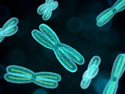
In the Chromosomes unit students learn the structure of the chromosome and identify the consequences of a base substitution mutation.
The unit is planned to take 3 school days
Essential idea:
- Chromosomes carry genes in a linear sequence that is shared by members of a species. 3.
Nature of science:
- Developments in research follow improvements in techniques—autoradiography was used to establish the length of DNA molecules in chromosomes. (1.8)
- Outline the advancement in knowledge gained from the development of autoradiography techniques.
- Outline the advancement in knowledge gained from the development of autoradiography techniques.
Understandings
3.2.U1 Prokaryotes have one chromosome consisting of a circular DNA molecule. (Oxford Biology Course Companion page 18).
- Describe the arrangement of prokaryotic DNA (nucleoid and plasmid).
- Define the term “naked” in relation to prokaryotic DNA
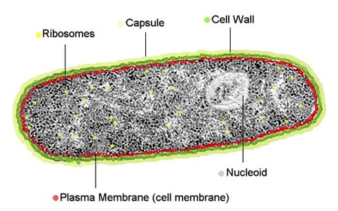
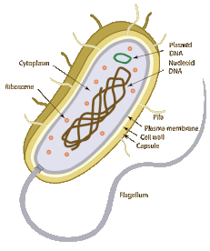
Prokaryotes do not possess a nucleus – instead genetic material is found free in the cytoplasm in a region called the nucleoid
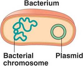
The genetic material of a prokaryote consists of a single chromosome consisting of a circular DNA molecule (genophore)
The DNA of prokaryotic cells is naked – meaning it is not associated with proteins for additional packagingProkaryotes have one chromosome consisting of a circular DNA molecule
The genetic material of a prokaryote consists of a single chromosome consisting of a circular DNA molecule (genophore)
The DNA of prokaryotic cells is naked – meaning it is not associated with proteins for additional packagingProkaryotes have one chromosome consisting of a circular DNA molecule
- Circular DNA molecule contains all the genes needed for the basic life processes of the cell
- DNA in bacteria is not related/does not work with proteins therefore is described as naked
- 1 chromosome is present in a prokaryotic cell, there is usually only a single copy of each gene, there are two identical copies briefly after the chromosome has been replicated but this is in preparation for cell division.
- the two identical chromosomes are moved to opposite poles and the cell then splits in two
3.2.U2 Some prokaryotes also have plasmids but eukaryotes do not. Oxford Biology Course Companion page 150).
- Describe the structure and function of plasmid DNA.
Plasmids are small, circular DNA molecules that contain only a few genes and are capable of self-replication. Plasmids are present in some prokaryotic cells, but are not naturally present in eukaryotic cells
- Plasmids are small extra DNA molecules that are found in prokaryotes, very unusual in eukaryotes
- Described as circular and naked, containing very few genes that are useful to the cell but are not needed for basic life processes
- Plasmids are not always formed at the same time as the chromosome of a prokaryotic cells or at the same rate, therefore there can be multiple copies of plasmids in a cell and it may not be passed to both cells formed by cell division
- Gene transfer between species – copies of plasmids can be transferred from one cell to another, allowing spread through a population. It is possible that the plasmids can cross through species barrier, happens when plasmid is released when a prokaryotic cell dies and is then absorbed by a cell of a different species
3.2.U3 Eukaryote chromosomes are linear DNA molecules associated with histone proteins (Oxford Biology Course Companion page 151).
- Describe the structure of eukaryotic DNA and associated histone proteins during interphase (chromatin).
- Explain why chromatin DNA in interphase is said to look like “beads on a string
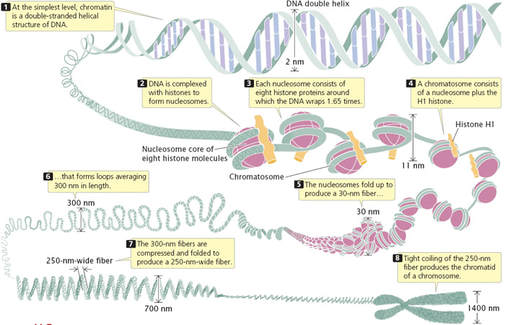
- Chromosomes in eukaryotes are made up of DNA/ protein
- DNA is a long/ single linear DNA molecules
- Related with histone proteins – which are globular in shape and wider than the DNA
- There are many of these histone molecules in a chromosome with DNA molecules wrapped around them
- Adjacent histones in the chromosome are separated by short stretches of the DNA molecule that are not in contact with histones – this is why the eukaryotic chromosome looks like a string of beads during interphase
3.2.U4 In a eukaryote species there are different chromosomes that carry different genes.
- List three ways in which the types of chromosomes within a single cell are different.
- State the number of nuclear chromosome types in a human cell.
Eukaryotic chromosomes are linear molecules of DNA that are compacted during cell division (mitosis or meiosis). Each chromosome has a constriction point called a centromere, which divides the chromosome into two sections (or ‘arms’). The shorter section is designated the p arm and the longer section is designated the q arm. The genetic material of eukaryotic cells consist of multiple linear molecules of DNA that are associated with histone proteins
The packaging of DNA with histone proteins results in a greatly compacted structure, allowing for more efficient storage
The packaging of DNA with histone proteins results in a greatly compacted structure, allowing for more efficient storage
- Eukaryotic chromosomes are too narrow to be visible with a light microscope during interphase –> then become visible during mitosis and meiosis because the chromosomes because shorter and fatter by supercoiling, they become visible only if stains that bind either DNA or proteins are used
- 1st stage of mitosis – chromosomes can be seen in double because there are two chromatids, that have identical DNA molecules produced by replication
- Chromosomes during mitosis can be seen differently whether they are different in length and/or in the position of the centromere where the two chromatids are held together
- in eukaryotes there are around two different types but in humans there are 23 different types of chromosomes
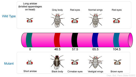
- Each gene in the eukaryotes has a different/certain position on one type of chromosome, called the locus of the gene
- Each chromosome type therefore carries a specific sequence of genes arranged along the linear DNA molecule – this can contain over a thousand genes
3.2.U5 Homologous chromosomes carry the same sequence of genes but not necessarily the same alleles of those genes. (Oxford Biology Course Companion page 152).
- Define homologous chromosome.
- State a similarity and a difference found between pairs of homologous chromosomes.
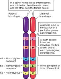
Sexually reproducing organisms inherit their genetic sequences from both parents . This means that these organisms will possess two copies of each chromosome (one of maternal origin ; one of paternal origin). These maternal and paternal chromosome pairs are called homologous chromosomes
Homologous chromosomes are chromosomes that share:
They are not identical to each other because some of the genes on them, the alleles are different
The species can be interbreed – if 2 eukaryotes are members of the same species we can expect each of the chromosomes in one of them to be homologous with at least one chromosome in the other.
Homologous chromosomes are chromosomes that share:
- The same structural features (e.g. same size, same banding patterns, same centromere positions)
- The same genes at the same loci positions (while the genes are the same, alleles may be different)homologous are two chromosomes that have the same sequence of genes
They are not identical to each other because some of the genes on them, the alleles are different
The species can be interbreed – if 2 eukaryotes are members of the same species we can expect each of the chromosomes in one of them to be homologous with at least one chromosome in the other.
3.2.U6 Diploid nuclei have pairs of homologous chromosomes. (Oxford Biology Course Companion page 155).
- Define diploid
- State the human cell diploid number
- Outline the formation of a diploid cell from two haploid gametes.
- State an advantage of being diploid.
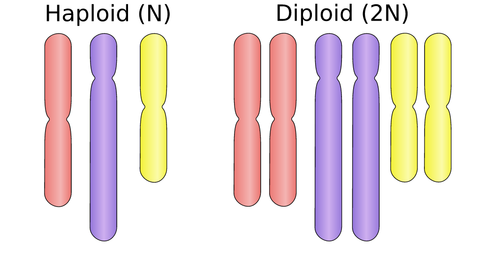
Nuclei possessing pairs of homologous chromosomes are diploid (symbolised by 2n)
- Two chromosomes of each type
- In humans that means it contains 46 chromosomes
- Haploid gametes fuse together during sex, a zygote with a diploid nucleus is formed through mitosis it divides and more cells with diploid nuclei are produced, many animals and plants only consist of diploid cells
- Diploid nuclei have two copies of every gene, apart from the sex chromosomes
- A benefit of this is that harmful recessive mutations can be avoided if a dominant alleles is also present.
- HYBRID VIGOUR – organism are more vigorous if they have two different alleles of genes instead of one
3.2.U7 Haploid nuclei have one chromosome of each pair. (Oxford Biology Course Companion page 154).
- Define haploid.
- State the human cell haploid number.
- List example haploid cells.
Nuclei possessing only one set of chromosomes are haploid (symbolised by n)
- They have one chromosome of each type
- Gametes – sex cells that fuse together during sexual reproduction
- gametes have haploid nuclei, so in humans both egg and sperm cells contain 23 chromosomes
3.2.U8 The number of chromosomes is a characteristic feature of members of a species. (Oxford Biology Course Companion page 155).
- State that chromosome number and type is a distinguishing characteristic of a species.
- List mechanisms by which a species chromosome number can change.
Chromosome number is a characteristic feature of members of a particular species. Organisms with different diploid numbers are unlikely to be able to interbreed (cannot form homologous pairs in zygotes)
- organisms with a different number of chromosomes are unlikely to be able to interbreed so all the interbreeding members of a species need to have the same number of chromosomes
- the number of chromosomes can change through time, if chromosomes become fused together or increase if splits occur
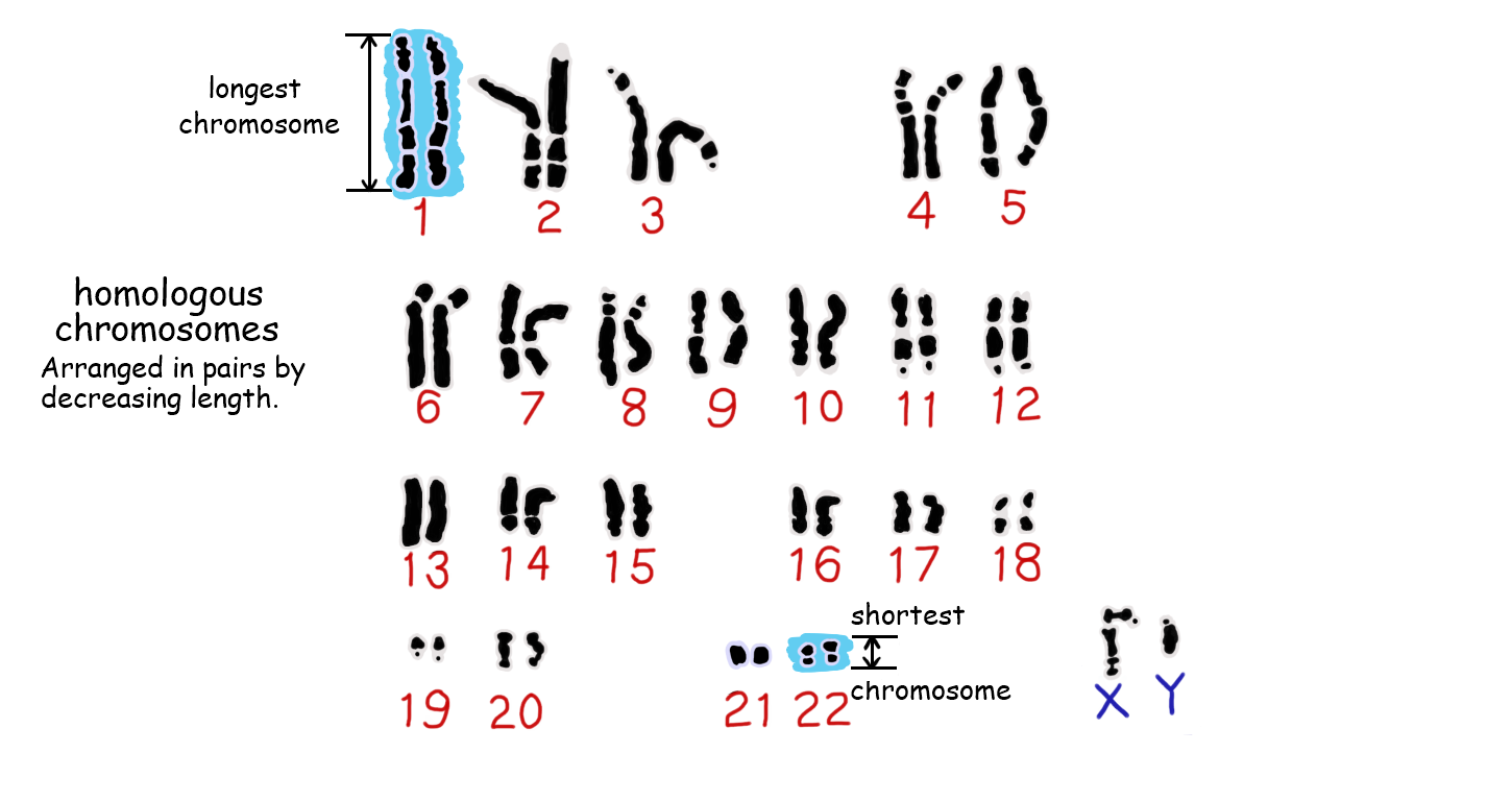
3.2.U9 A karyogram shows the chromosomes of an organism in homologous pairs of decreasing length. (Oxford Biology Course Companion page 157).
- Describe the process of creating a karyogram.
- List the characteristics by which chromosomes are arranged on the karyogram.
Karyotypes are the number and types of chromosomes in a eukaryotic cell – they are determined via a process that involves:
- Harvesting cells (usually from a foetus or white blood cells of adults)
- Chemically inducing cell division, then arresting mitosis while the chromosomes are condensed
- The stage during which mitosis is halted will determine whether chromosomes appear with sister chromatids or not. Taken during mitosis – cells in metaphase. The chromosomes of an organism are arranged into homologous pairs according to size (with sex chromosomes shown last)
- The chromosomes are stained and photographed to generate a visual profile that is known as a karyogram– distinctive banding pattern
- Stain dividing cells→ placed on microscope → burst by pressing → micrograph
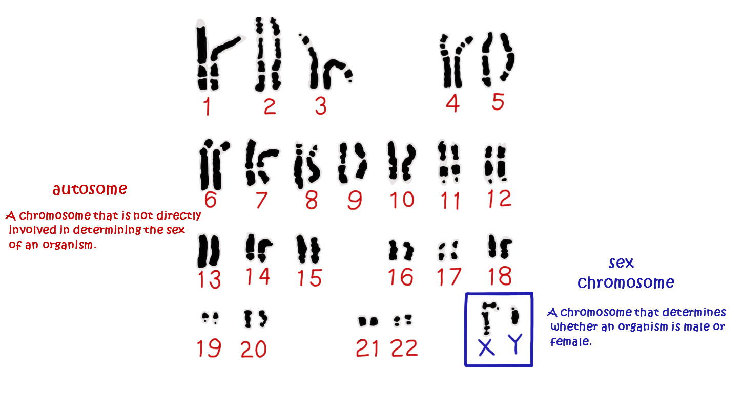
3.2.U10 Sex is determined by sex chromosomes and autosomes are chromosomes that do not determine sex.(Oxford Biology Course Companion page 157).
- Outline the structure and function of the two human sex chromosomes.
- Outline gender determination by sex chromosomes.
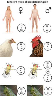 image from Wikipedia
Human sex determination occurs according to the X - Y system
- Females have two copies of the larger X chromosome
- Males have one X and one Y chromosome (and hence determine gender in offspring)
- The x chromosome is pretty big and has its centromere near the middle (women)
- y chromosome is much smaller and has its centromere near the end
- x has many genes that are important to both genders
- y only has a small number of genes, a small number of the y chromosomes has the same sequence of genes as a small part of the x chromosome, but the genes on the remainder of the y chromosome are not found on the x chromosome and are not needed for female development
- Fetus with 1 x and 1 y becomes a male, fetus with 2 x and 0 y becomes a female
Application
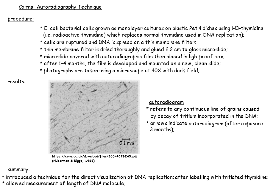
3.2.A1 Cairns’ technique for measuring the length of DNA molecules by autoradiography. (Oxford Biology Course Companion page 151).
- Describe Cairn’s technique for producing images of DNA molecules from E. coli.
- Outline conclusions drawn from the images produced using Cairn’s autoradiography technique.
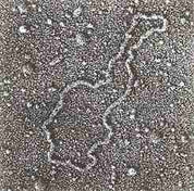
John Cairns pioneered a technique for measuring the length of DNA molecules by autoradiography
Previously, chromosome length could only be measured while condensed during mitosis (very inaccurate due to supercoiling). Cairns used autoradiography to visualise the chromosomes whilst uncoiled, allowing for more accurate indications of length. By using tritiated uracil (3H-U), regions of active transcription can be identified within the uncoiled chromosome
Previously, chromosome length could only be measured while condensed during mitosis (very inaccurate due to supercoiling). Cairns used autoradiography to visualise the chromosomes whilst uncoiled, allowing for more accurate indications of length. By using tritiated uracil (3H-U), regions of active transcription can be identified within the uncoiled chromosome
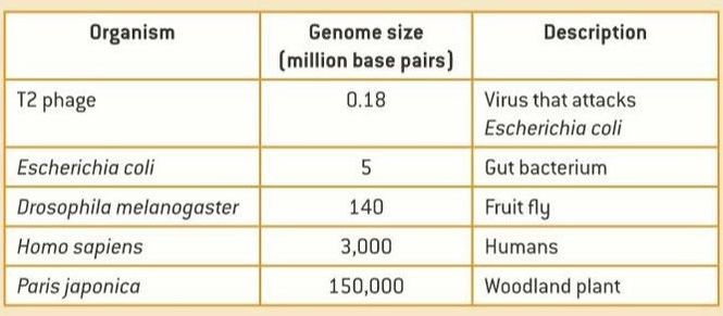
3.2.A2 Comparison of genome size in T2 phage, Escherichia coli, Drosophila melanogaster, Homo sapiens and Paris japonica.
Genome size can vary greatly between organisms and is not a valid indicator of genetic complexity. The largest known genome is possessed by the canopy plant Paris japonica – 150 billion base pairs. The smallest known genome is possessed by the bacterium Carsonella ruddi – 160,000 base pairs.
As a general rule:
As a general rule:
- Viruses and bacteria tend to have very small genomes
- Prokaryotes typically have smaller genomes than eukaryotes
- Sizes of plant genomes can vary dramatically due to the capacity for plant species to self-fertilise and become polyploid
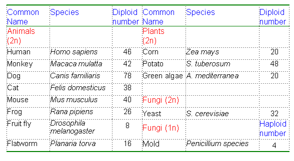
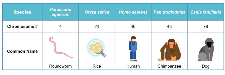
3.2.A3 Comparison of diploid chromosome numbers of Homo sapiens, Pan troglodytes, Canis familiaris, Oryza sativa, Parascaris equorum. (Oxford Biology Course Companion page 155).
- State the minimum chromosome number in eukaryotes.
- Explain why the typical number of chromosomes in a species is always an even number.
- Explain why the chromosome number of a species does not indicate the number of genes in the species.
- Explain the relationship between the number of human and chimpanzee chromosomes.
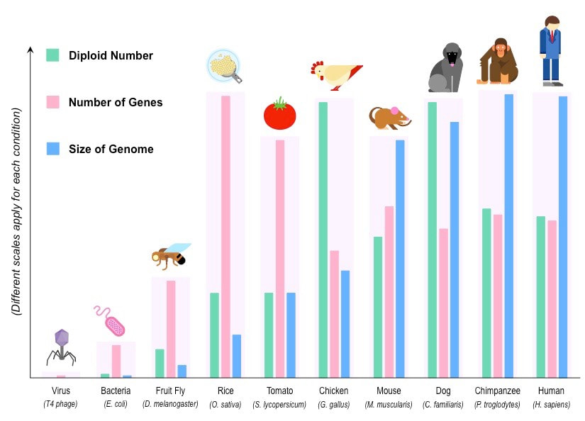
Chromosome number does not provide a valid indication of genetic complexity, for instance:
- Tomatoes (Solanum lycopersicum) have 24 chromosomes and a genome size of 950 million bp, but possess ~32,000 genes
- Chickens (Gallus gallus) have 78 chromosomes and a genome size of 1.2 billion bp, but possess only ~17,000 genes
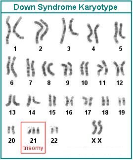
3.2.A4 Use of karyograms to deduce sex and diagnose Down syndrome in humans (Oxford Biology Course Companion page 158).
- Distinguish between a karyogram and a karyotype.
- Deduce the sex of an individual given a karyogram.
- Describe the use of a karyogram to diagnose Down syndrome.
Karyotyping will typically occur prenatally and is used to:
Down syndrome is a condition whereby the individual has three copies of chromosome 21. It is caused by a non-disjunction event in one of the parental gametes. The extra genetic material causes mental and physical delays in the way the child develops
- Determine the gender of the unborn child (via identification of the sex chromosomes)
- Test for chromosomal abnormalities (e.g. aneuploidies or translocations)
Down syndrome is a condition whereby the individual has three copies of chromosome 21. It is caused by a non-disjunction event in one of the parental gametes. The extra genetic material causes mental and physical delays in the way the child develops
Skills:
3.2.S1 Use of databases to identify the locus of a human gene and its polypeptide product.
The locus of a human gene and its polypeptide product can both be identified using a single online resource:
GenBank – a genetic database that serves as an annotated collection of DNA sequences
dentifying Gene Loci
GenBank can be used to identify the specific location of a gene on any given chromosome
To identify a specific gene locus:
Identifying Polypeptide Products:
To identify the polypeptide product of a gene:
GenBank – a genetic database that serves as an annotated collection of DNA sequences
dentifying Gene Loci
GenBank can be used to identify the specific location of a gene on any given chromosome
To identify a specific gene locus:
- Change the search parameter from nucleotide to gene and type in the name of the gene of interest
- Choose the species of interest (i.e. Homo sapiens) and click on the link (under ‘Name / Gene ID’)
- Scroll to the ‘Genomic context’ section to determine the specific position of the gene locus
- A visual profile can be generated by clicking on ‘Map Viewer’ link and looking at the Ideogram on the left side
Identifying Polypeptide Products:
- GenBank can also be used to identify the polypeptide product of any given gene
To identify the polypeptide product of a gene:
- Change the search parameter from nucleotide to gene and type in the name of the gene of interest
- Choose the species of interest (i.e. Homo sapiens) and click on the link (under ‘Name / Gene ID’)
- The polypeptide product should be identified within the ‘Summary’ section
Key Words:
|
chromosomes
allele autosomes autoradiography chromatid staining Cairn sex chromosomes Paris japonica circular DNA Down syndrome |
gene
genome mutation base deletion centromere circular DNA prokaryotic autoradiography Canis family hitsone |
diploid cells
Down syndrome sequence haploid cells gene nuclei nucleoid T2 phage Oryza sativa Y chromosome |
chistone
homologous chromosomes karyogram karyotype loci Non-sister chromatids chromatin E. coli P. equorum sex determination |
naked DNA
plasmid sex chromosomes sister chromosome autosome autoradiography karyogram Drosophila karyotype X chromosome |
Class Assignment:
How Genes Work
Genome Sizes worksheet
Human Genome Project activity
Sickle Cell Anemia project
Sickle Cell Anemia poster
Mutation worksheet
MOLO mutation activity
Topic 3.2 Review
How Genes Work
Genome Sizes worksheet
Human Genome Project activity
Sickle Cell Anemia project
Sickle Cell Anemia poster
Mutation worksheet
MOLO mutation activity
Topic 3.2 Review
Powerpoint and notes on Topic 3.2 by Chris Payne
Your browser does not support viewing this document. Click here to download the document.
Your browser does not support viewing this document. Click here to download the document.
Correct use of terminology is a key skill in Biology. It is essential to use key terms correctly when communicating your understanding, particularly in assessments. Use the quizlet flashcards or other tools such as learn, scatter, space race, speller and test to help you master the vocabulary.
External Links:
Let’s start with a tour of the basics from Learn.Genetics at Utah.
DNA Coiling To Form Chromosomes
Chromosome Description
Video Tour of Basic Genetics
Click here for additional information on HOW GENES WORK.
Click here for some more information on Sickle Cell
DNA coiling on histone proteins from biostudio.com
A description of chromosomes from Dexter Pratt
Zooming in to Chromosome 11 (a bit too advanced) from the DNA Learning Centre’s Gene Almanac
Transcription Java game from thinkquest.org
How do mutations occur? from the DNAi at the Dolan DNA Learning Centre
In the News:
Scientists Finally Pronounce Human Genome (2013-08-15, satire)
Why Are Some People Left-Handed? (2013-09-12)
Increased Height in HFE Hemochromatosis (2013-08-22)
International-mindedness:
- Sequencing of the rice genome involved cooperation between biologists in 10 countries.
Video Clips:
Animated and narrated segments presenting all the essential steps in sequencing a genome. From the NHGRI's Online Education Kit: Understanding the Human Genome Project.
Evolution of Sickle Cell: Resistance to Malaria
Epigenetics: A new frontier in heredity
