Topic 11.2: Movement
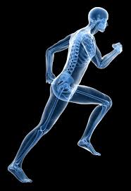 image from Better Movement
In the Movement unit we will learn that muscles allow us to move. Smooth muscle and cardiac muscle move to facilitate body functions like heartbeats and digestion. We will also learn the movement of these muscles is directed by the autonomic part of the nervous system—those are the nerves that control organs. Skeletal muscles move our bodies in space. They take direct instruction from the specific nerves that innervate each muscle. Want to learn more about the muscles in the human body? Here are five other facts to keep in mind about the muscular system.
This unit will last 4 school days.
This unit will last 4 school days.
Essential idea:
- The roles of the musculoskeletal system are movement, support and protection.
Nature of science:
- Developments in scientific research follow improvements in apparatus—fluorescent calcium ions have been used to study the cyclic interactions in muscle contraction. (1.8)
Understandings
11.2 U 1 Bones and exoskeletons provide anchorage for muscles and act as levers. (Oxford Biology Course Companion page 476)
- State the function of bones and exoskeletons.
- Contrast bones with exoskeletons.
- Identify the fulcrum, effort force and resultant force in the motion of the spine and the grasshopper leg.
- Determine the class of motion of a lever
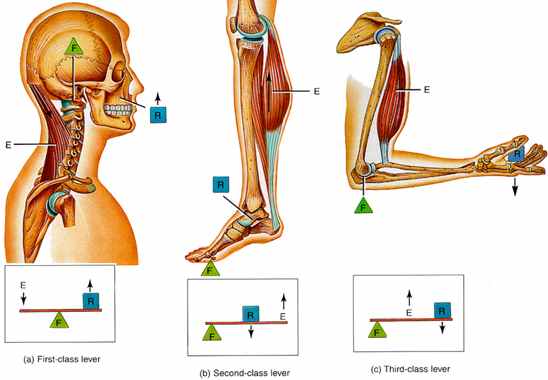
Muscles and bones act together to form levers. A lever is a rigid rod (usually a length of bone) that turns about a pivot (usually a joint). Levers can be used so that a small force can move a much bigger force. This is called mechanical advantage.
Levers can be used so that a small force can move a much bigger force. This is called mechanical advantage. In our bodies bones act as lever arms, joints act as pivots, and muscles provide the effort forces to move loads
- Bones act as levers so the body can move and provide structural support (skeleton).
- Ligaments are strong bands that connect bone to bone strengthening the joint during movement.
- Tendons have dense connective tissue that connects muscles to bones, allowing movement of the bone when a muscle contracts.
- Muscles provide the force for movement by contracting (shortens the muscle fibers)
- The joint acts as a pivot point or a fulcrum
- The force applied (when the muscle contracts) is called the effort
- The force or load needed to overcome for movement to take place is called the resistance
Levers can be used so that a small force can move a much bigger force. This is called mechanical advantage. In our bodies bones act as lever arms, joints act as pivots, and muscles provide the effort forces to move loads
- Levers are classified by first, second, and third class, depending upon the positions among the fulcrum, the effort, and the resistance.
- First-class levers have the fulcrum in the middle, like a seesaw. An example of a first class lever is when a human nods their head (top of the spinal column is the fulcrum, the effort force is provided by the muscles in the back of the neck, and the resistance is weight of the head).
- Second-class levers have a resistance in the middle, like a load in a wheel-barrow. The body acts as second class lever when engaged in push up or calf raise. During a calf raise ball of foot is fulcrum, the body’s mass is the resistance and the effort is applied by calf muscle.
- Third-class levers have the effort from the muscle in the middle of the lever. The majority of the human body's musculoskeletal levers are third class. These levers are built for speed and range of motion. Muscle attachments are usually close to the fulcrum. In the example of the arm, the effort force is provided by the contraction of the biceps, the fulcrum is the elbow joint and the resistance would be provided by whatever weight is being lifted.
- Exoskeletons in insects and crustaceans can facilitate the movement by providing an anchorage for muscles; similarly to how bones provide anchorage for animals with internal skeletons.
11.2 U 2 Synovial joints allow certain movements but not others. (Oxford Biology Course Companion page 477).
- Define antagonistic pairs in relation to muscle movement.
- State an example of an antagonistic pair of muscles.
The type of joint determines the amount of movement that is possible. For ball and socket joints, such as the hip or the shoulder, movement through all three planes are possible. At the hip joint, the head of the femur is the ball the fits into the socket of the pelvis. The movements possible at the joint are flexion, extension, rotation, abduction and adduction.. For hinge joints, such as the knee, flexions (bending) and extensions (straightening) are the possible movements (movement in one plane); however, slight side to side movements are possible.
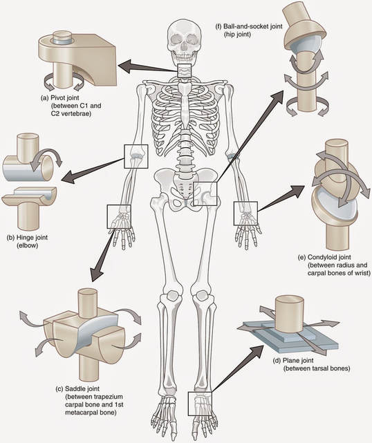
image from wikipedia
|
|
|
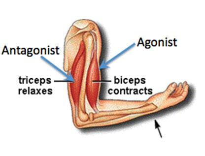
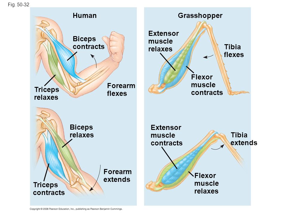
11.2 U 3 Movement of the body requires muscles to work in antagonistic pairs. (Oxford Biology Course Companion page 479).
- Compare the motion of hinge joints with the motion of a ball and socket joint.
- Outline motion of the human knee, shoulder and hip in terms of flexion, extension, rotation, abduction and adduction.
Skeletal muscles occurs in pairs that are antagonistic. This means that when one contracts, the other relaxes. Antagonistic muscles produce opposite movements at a joint. For example, in the elbow, the triceps extends the forearm while the biceps flex the forearm.
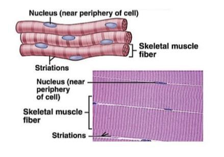
11.2 U 4 Skeletal muscle fibres are multinucleate and contain specialized endoplasmic reticulum. (Oxford Biology Course Companion page 480).
- List three types of muscle tissue found in the human body.
- Label a diagram of a muscle fibre cell, including the sacrolemma, nuclei, sacroplasmic reticulum and mitochondria.
A whole skeletal muscle is considered an organ of the muscular system. Each organ or muscle consists of skeletal muscle tissue, connective tissue, nerve tissue, and blood or vascular tissue. Skeletal muscles vary considerably in size, shape, and arrangement of fibers. They range from extremely tiny strands such as the stapedium muscle of the middle ear to large masses such as the muscles of the thigh. Some skeletal muscles are broad in shape and some narrow. In some muscles the fibers are parallel to the long axis of the muscle; in some they converge to a narrow attachment; and in some they are oblique.
The 3 types of muscle tissue are cardiac, smooth, and skeletal. Cardiac muscle cells are located in the walls of the heart, appear striated, and are under involuntary control. Smooth muscle fibers are located in walls of hollow visceral organs, except the heart, appear spindle-shaped, and are also under involuntary control. Skeletal muscle fibers occur in muscles which are attached to the skeleton. They are striated in appearance and are under voluntary control.
The 3 types of muscle tissue are cardiac, smooth, and skeletal. Cardiac muscle cells are located in the walls of the heart, appear striated, and are under involuntary control. Smooth muscle fibers are located in walls of hollow visceral organs, except the heart, appear spindle-shaped, and are also under involuntary control. Skeletal muscle fibers occur in muscles which are attached to the skeleton. They are striated in appearance and are under voluntary control.
- Skeletal muscles are composed of bundles of muscle fibers and have a striped appearance because of areas of thick and thin filaments (myosin and actin)
- Muscle cells have many nuclei and are long because the embryonic muscle cells fuse together
- Muscle fibers are composed of many parallel elongated fibers called myofibrils
- A modified endoplasmic reticulum, called the sarcoplasmic reticulum (fluid-filled membranous sacs), extends throughout the muscle fibre, wrapping around each myofibril, sending a signal to the all parts of the muscle fibre to contract at the same time
- The sarcoplasmic reticulum stores calcium - between myofibrils are large numbers of mitochondria, which provide ATP needed for contractions.
11.2 U 5 Muscle fibres contain many myofibrils. (Oxford Biology Course Companion page 480).
- Outline the relationship between muscles, muscle fibre cells and myofibrils
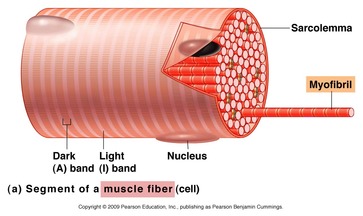
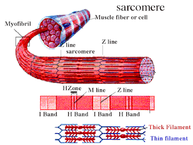
A skeletal muscle is made up of many multinucleate muscle fibers (cells), each of which runs the entire length of the muscle and is connected to at least one motor neuron. Each muscle fiber contains multiple bundles of contractile protein called myofibrils. Myofibrils made up units called sacromere, strung out in single file along the length of myofibrils.
Within each muscle fibre there are many parallel, elongated structures called myofibrils. These have alternating light and dark bands, which give striated muscle its strips. In the centre of each light band is a disc-shaped structure, referred to as the Z-line.
Within each muscle fibre there are many parallel, elongated structures called myofibrils. These have alternating light and dark bands, which give striated muscle its strips. In the centre of each light band is a disc-shaped structure, referred to as the Z-line.
11.2 U 6 Each myofibril is made up of contractile sarcomeres. (Oxford Biology Course Companion page 481).
- Outline the relationship between myofibrils and sacromeres.
- Describe a structure of a sarcomere., including the Z line, thin actin filaments, thick myosin filaments, light band and dark band.
Myofibrils – rod-shaped parallel bodies consisting of actin and myosin filaments
Sarcomere
- Sarcolemma – plasma membrane of the muscle cell
- Mitochondria – large numbers; found dispersed around individual myofibrils
Sarcomere
- Lies between two Z-lines which are dense protein discs
- Contains the thick filament (myosin) and thin filament (actin)
- Myosin contains a head which binds to the binding site on the actin; interaction between myosin and actin (cross-bridge) is responsible for muscle contraction
- Myosin is seen as dark bands while actin is seen as light bands
- Actin filaments are attached to a Z-line at one end
- Myosin filaments are interdigitated with actin filaments at both ends and occupy the centre of the sarcomere
- Each mysosin filament is surrounded by six actin filaments
- A bands contain a full length of myosin and some of the actin filaments
- I bands contain only actin filaments
11.2 U 7 The contraction of the skeletal muscle is achieved by the sliding of actin and myosin filaments. (Oxford Biology Course Companion page 482)
- Explain the sliding-filament mechanism of muscle contraction, including the role of myosin heads, cross bridges and ATP
During a muscle contraction, myosin filaments pull actin filaments towards the centre of the sarcomere. This shortens the sarcomere and the overall length of the muscle fibre. When this occurs, the myosin heads bind to sites on the actin filaments, creating cross-bridges, pulling (sliding) the actin filaments along the myosin filaments with energy from ATP . This is called sliding filament theory and is explained further below
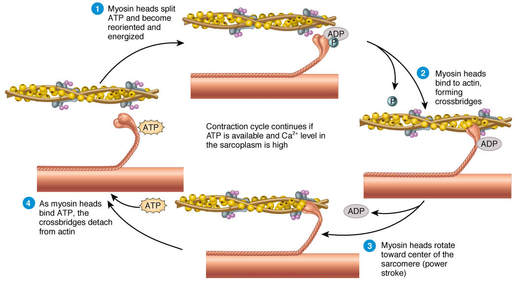
image from Fitness Acadamey
11.2 U 8 ATP hydrolysis and cross bridge formation are necessary for the filaments to slide. (Oxford Biology Course Companion page 483).
- Explain the exposure of myosin head binding sites on actin, including the role of the sarcoplasmic reticulum, calcium, troponin and tropomyosin.
- ATP attaches to the myosin heads breaking the cross-bridges between the myosin heads and actin binding sites
- The ATP undergoes a hydrolysis reaction forming ADP + Pi.
- This causes a positional change in the myosin head (cocked back).
- The myosin heads bind to actin filaments forming cross-bridges at a site one position further from the centre of the sarcomere
- When the ADP + Pi are released the myosin heads change conformational position, sliding the actin filaments towards the center of the Sarcomere.
- This is called the “power stroke”.
- After the power stroke ATP again binds to the myosin head, causing it to detach from the actin filament ready for another cycle.
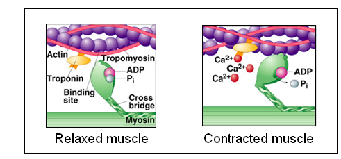
11.2 U 9 Calcium ions and the proteins tropomyosin and troponin control muscle contractions. (Oxford Biology Course Companion page 483)
- List the events that occur during cross-bridge cycles.
- Describe the role of ATP in muscle contraction.
- An action potential propagated along a motor neuron arrives at the neuromuscular junction.
- This causes the release of the neurotransmitter acetylcholine into the synapse between the terminal axon of the motor neuron and the sarcolemma of the skeletal muscle.
- The acetylcholine binds to receptors on the sarcolemma, causing voltage-gated channels to open and Na+ ions to flow into the muscle cells.
- This creates an action potential in the striated muscle.
- The action potential is further propagated along the sarcolemma of the skeletal muscle.
- The action potential moves into the interior of the muscle cell through folds called t tubules.
- The depolarization of the t tubules causes voltage-gated Ca+ channels on the sarcoplasmic reticulum to open, causing an influx of Ca+ ions into the sarcoplasm.
- Ca+ ions bind to troponin which causes tropomyosin to move exposing the myosin binding sites (troponin and tropomyosin are regulatory proteins blocking the myosin binding sites).
Applications
11.2 A 1 Antagonistic pairs of muscles in an insect leg (Oxford Biology Course Companion page 478).
- Label the tibia, femur, tarsus, flexor muscle and extensor muscle on a diagram of a grasshopper hindlimb.
- Describe the contraction of muscles and movement of hindlimb structures that produces a grasshopper jump.
Grasshoppers
- The hind limbs of grasshoppers are specialized for jumping
- It has a jointed appendage with three parts
- Below the joint is the tibia, and at the base of the tibia is another joint called the tarsus
- Above the joint is the femur
- When the grasshopper jumps, the flexor muscles contract, and the femur and tibia are brought closer together (flexing)(extensor muscles are relaxed)
- As the grasshopper jumps the extensor muscles contract, extending the tibia, creating a powerful jump force
Skills
11.2 S 1 Annotation of a diagram of the human elbow. include cartilage, synovial fluid, joint capsule, named bones and named antagonistic muscles. (Oxford Biology Course Companion page 478).
- Label a diagram of the human elbow inclusive of: humerus, triceps, biceps, joint capsule, synovial fluid, radius, cartilage and ulna.
- State the function of structures found in the human elbow, including: humerus, triceps, biceps, joint capsule, synovial fluid, radius, cartilage and ulna.
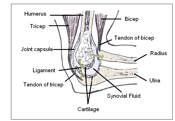
image from MONARDO.INFO

11.2 S 2 Drawing labelled diagrams of the structure of a sarcomere. Include Z lines, actin filaments, myosin filaments with heads, and the resultant light and dark bands. (Oxford Biology Course Companion page 481).
- Draw a diagram of the structure of a sarcomere.
- Label a sarcomere diagram, including Z lines, actin filaments, myosin filaments with heads and the resultant light and dark bands.
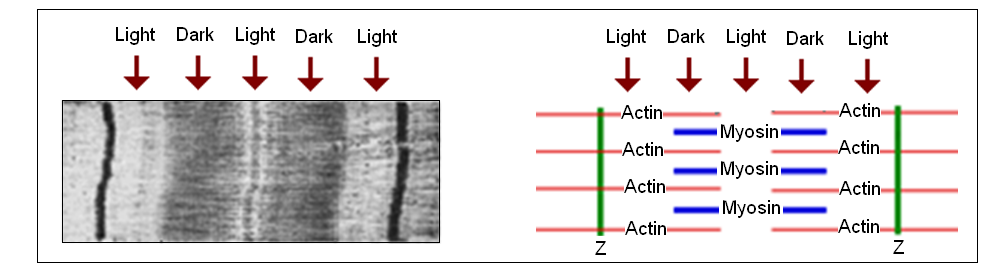
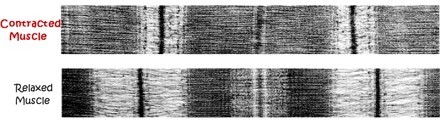
11.2 S 3 Analysis of electron micrographs to find the state of contraction of muscle fibres. Measurement of the length of sarcomeres will require calibration of the eyepiece scale of the microscope. (Oxford Biology Course Companion page 482)
- Compare a relaxed sarcomere to a contracted sarcomere, referring to Z line distance and size of light bands.
- Determine of a sarcomere is contracted or relaxed given an electron micrograph image.
Notice in the fully contracted sarcomere the actin filaments slide along the myosin causing the light bands to shorten, even though the dark bands stay the same length. The Z lines get closer together as the sarcomere contracts.. The muscle also can be in various states of partial contraction.
Key Terms
|
musculoskeletal
effort force ball joint nuclei thick myosin filaments myosin heads troponin extensor muscle radius bioluminescence flexion |
exoskeletons
resultant force synovial joints Z-line light band cross bridges tibia hindlimb cartilage 1st class lever abduction |
muscles
lever muscle fibers myofibrils dark band ATP femur triceps ulna scapula adduction |
bone
antagonistic pairs endoplasmic reticulum sacroplasmic reticulum thin actin filaments calcium tarsus biceps sarcomere 2nd class lever rotation |
fulcrum
hinge joint sacrolemma mitochondria sliding-filament tropomyosin flexor muscle joint capsule, synovial fluid 3rd class lever |
PowerPoint and Notes on Topic 11.2 by Chris Payne
Correct use of terminology is a key skill in Biology. It is essential to use key terms correctly when communicating your understanding, particularly in assessments. Use the quizlet flashcards or other tools such as learn, scatter, space race, speller and test to help you master the vocabulary.
How Muscles Work - Science Daily, Aug 2017
Scientists find power switch for muscles - Science Daily, Mar 2018
Scientists find power switch for muscles - Science Daily, Mar 2018
Video Clips
Movement of the body's skeleton is made possible by a number of body parts working together including the joints. A hinge joint at the elbow allows the arm to bend and extend. The condylar joint at the knee gives similar movement plus some rotation. Mobile ellipsoid joints between the fingers and palm of the hand allow circular movements but no rotation. Ball-and-socket joints, like the one at the shoulder, give maximum freedom of movement. At the other extreme, plane joints between the toe bones permit only a small degree of gliding movement and the pivot joint between the two cervical vertebrae-the atlas and the axis-merely provides rotation of the head. The saddle joint at the ankle is almost as mobile as the shoulder joint, but rotation is far more limited.
Each time you take a step, 200 muscles work in unison to lift your foot, propel it forward, and set it down. It’s just one of the many thousands of tasks performed by the muscular system: this network of over 650 muscles covers the body and is the reason we can blink, smile, run, jump, and stand upright. So how does it work?
We continue our look at your bones and skeletal system, skipping over the silly kid's song in favor of a more detailed look at your your axial and appendicular skeleton. This episode also talks about the structural and functional classifications of your joints and the major types of body movement that they facilitate.
We're kicking off our exploration of muscles with a look at the complex and important relationship between actin and myosin. Your smooth, cardiac, and skeletal muscles create movement by contracting and releasing in a process called the sliding filament model. Your skeletal muscles are constructed like a rope made of bundles of protein fibers, and that the smallest strands are your actin and myosin myofilaments. Its their use of calcium and ATP that causes the binding and unbinding that makes sarcomeres contract and relax.
Hank calls in a friend to do his push ups for him today to explain how skeletal muscles work together to create and reverse movements. Hank and Claire also demonstrate the role size plays in motor units, the three phase cycle of muscle twitches, and how the strength and frequency of an impulse affects the strength and duration of a contraction. This episode also explains twitch summation, tetanus, and isotonic vs. isometric movements.
Impulse to activate action potential in skeletal muscle,
Muscle contraction animation
