topic 6.5: neurones and Synapses
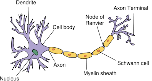
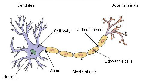
In the Neurons and Synapses unit you will learn that the nervous system consists of the central nervous system (CNS) and the peripheral nerves. It is composed of cells called neurons that can carry rapid electrical impulses. There are two types of neurons: sensory neurons which carry nerve impulses from sense organs to the central nervous system and motor neurons which carry nerve impulses from the central nervous system to effectors which produce the response. Within the CNS the impulses are carried by relay neurons. Figure 1 below illustrates a structure of the motor neuron.
This unit will last 3 school days.
This unit will last 3 school days.
Essential idea:
- Neurons transmit the message, synapses modulate the message.
Nature of science:
The Centre for Neural Circuits and Behaviour at Oxford University is an excellent example of collaboration between scientists with different areas of expertise. The four group leaders of the research team and the area of science that they originally studied are:
source: Oxford University Press IB Course Companion
- Cooperation and collaboration between groups of scientists—biologists are contributing to research into memory and learning. (4.3)
The Centre for Neural Circuits and Behaviour at Oxford University is an excellent example of collaboration between scientists with different areas of expertise. The four group leaders of the research team and the area of science that they originally studied are:
- Professor Gero Miesenböck – medicine and physiology
- Dr Martin Booth – engineering and optical microscopy
- Dr Korneel Hens – chemistry and biochemistry
- Professor Scott Waddell – genetics, molecular biology and neurobiology.
source: Oxford University Press IB Course Companion
Understandings:
6.5.U1 Neurons transmit electrical impulses [The details of structure of different types of neuron are not needed.] (Oxford Biology Course Companion page 319)
- State the function of the nervous system.
- Draw the structure of a neuron.
- Annotate a neuron drawing with the name and function of the following cell parts: dendrites, axon and cell body
One form of internal communication in the body occurs through nerve impulses in the nervous system. Neurons transmit electrical impulses by allowing the passage of charged ions across their membranes in response to stimuli. . While neurons may differ according to role (sensory, relay or motor), most share three basic components:
- Dendrites – Short-branched fibres that convert chemical information from other neurons or receptor cells into electrical signals
- Axon – An elongated fibre that transmits electrical signals to terminal regions for communication with other neurons or effectors
- Soma – A cell body containing the nucleus and organelles, where essential metabolic processes occur to maintain cell survival
6.5.U2 The myelination of nerve fibres allows for saltatory conduction. (Oxford Biology Course Companion page 3200
- Outline the structure and function of myelin.
- State the role of Schwann cells in formation of myelin.
- Outline the mechanism and benefit of saltatory conduction.
- Compare the speed of nerve impulse conduction myelinated and non-myelinated neurons.
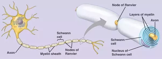
Nerve fibres conduct electrical impulses along the length of their axons. Some of these axons such as interneurons are unmyelinated, and therefore the impulse travels much slower. Some axons are surrounded by a mixture of protein and phospholipids called myelin that collectively form a myelin sheath. The greater the diameter, the greater the speed of the nerve impulse.
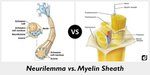
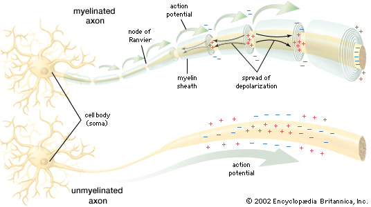
Many layers of myelin are deposited around the axon by special cells called Schwann cells. In between the myelin are gaps called the nodes of Ranvier. In myelinated neurons, the impulse can jump from one node to the next. This is called saltatory conduction. This allows myelinated neurons to conduct impulses up to 100x faster than unmyelinated axons
- Myelin is a mixture of protein and phospholipids that is produced by glial cells (Schwann cells in PNS; oligodendrocytes in CNS)
Many layers of myelin are deposited around the axon by special cells called Schwann cells. In between the myelin are gaps called the nodes of Ranvier. In myelinated neurons, the impulse can jump from one node to the next. This is called saltatory conduction. This allows myelinated neurons to conduct impulses up to 100x faster than unmyelinated axons
Nerve fibers and saltatory conduction
6.5.U3 Neurons pump sodium and potassium ions across their membranes to generate a resting potential. (Oxford Biology Course Companion page 321)
- Define resting potential.
- Explain three mechanisms that together create the resting potential in a neuron.
- State the voltage of the resting potential.
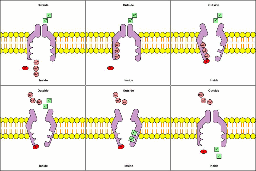
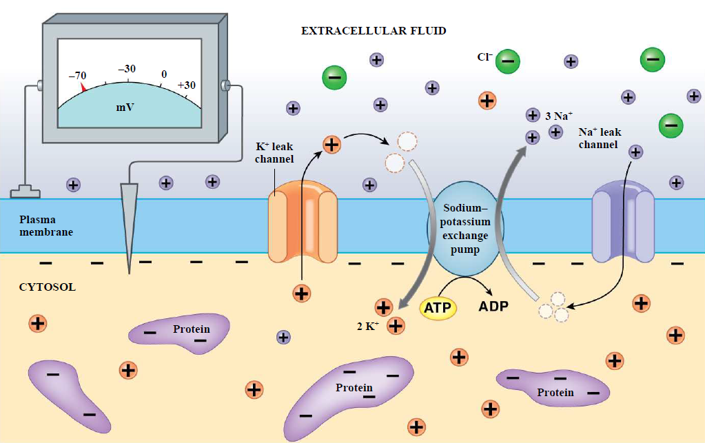
When a neuron is inactive, there is an imbalance of positive and negative charges across its membrane. This results in a potential called the resting potential. To create this potential, sodium-potassium pumps transfers Na+ ions out of the cell and K+ ions in with a 3:2 ratio. This creates a charge imbalance across the membrane of approximately -70 mV.
Neurons generate and conduct electrical signals by pumping positively charged ions (Na+ and K+) across their membrane
Neurons generate and conduct electrical signals by pumping positively charged ions (Na+ and K+) across their membrane
- The time period when a neuron that is not conducting a nerve impulse, but is ready to conduct one, is called the resting potential.
- This membrane potential is due to an imbalance of positive and negative charges across the membrane
- Sodium-potassium pumps pump Na+ out of the axon and K+ into the axon
- Three Na+ are pumped out of the neuron and two K+ are pumped into the neuron
- This creates a concentration gradient of Na+ (outside to in) and of K+ (inside to out)
- The membrane is also much more permeable to K+ as Na+, so K+ leaks back out of the neuron through leak channels
- This means the Na+ concentration is much greater outside the neuron
- There are also negatively charged ions permanently located in the cytoplasm of the neuron
- These conditions create a resting membrane potential of -70 mV inside the neuron
6.5.U4 An action potential consists of depolarization and repolarization of the neuron. (Oxford Biology Course Companion page 321)
- Define action potential, depolarization and repolarization.
- Outline the mechanism of neuron depolarization.
- Outline the mechanism of neuron repolarization
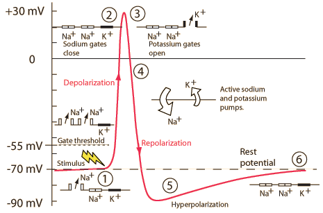
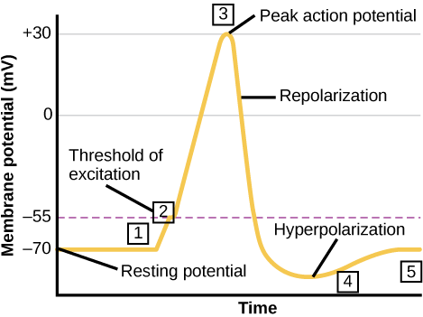
Action potentials are the rapid changes in charge across the membrane that occur when a neuron is firing. Action potentials occur in three main stages: depolarization, repolarization and a refractory period
- Action potentials are rapid changes in membrane potentials
- This consists of a rapid depolarization (change from negative to positive when sodium diffuses into the neuron) and a rapid repolarization (change from positive to negative when potassium diffuse out of the neuron)
- The arrival of an action potential caused by a stimulus causes a depolarization of the membrane as Na+ channels begin to open.
- If the membrane potential reaches a threshold level of -50mV. Many more voltage-gated Na+ channels open and Na+ rapidly diffuses into the neuron
- The inside of the neuron becomes more positively charged than the outside of the neuron (depolarization)
- K+ channels open and K+ ions diffuse out of the neuron making the inside negative again (repolarization)
- After the action potential, there is a refractory period where the impulse cannot go back in the same direction. This ensures a one-way nerve impulse
6.5.U5 Nerve impulses are action potentials propagated along the axons of neurons. (Oxford Biology Course Companion page 322)
- Define nerve impulse.
- Describe how nerve impulses are propagated along the neuron axon.
- Outline the cause and consequence of the refractory period after depolarization.
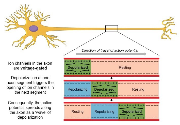
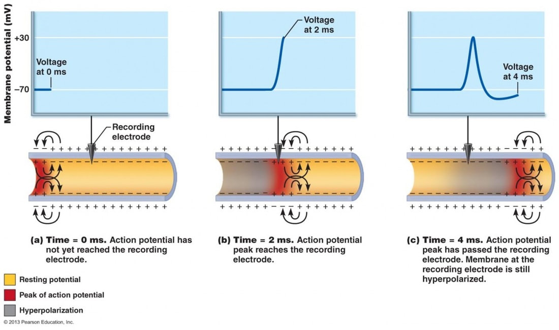
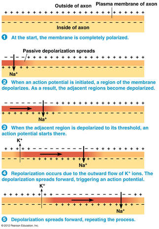
Nerve impulses are action potentials that move along the length of an axon as a wave of depolarisation
- As a depolarization occurs in one part of the neuron, the positive charge triggers the Na+ channels to open in the nearby regions causing an action potential to occur.
- This action potential will cause a depolarization in the next region.
- The propagation of action potentials will continue along the axon of the neuron.
- Nerve impulses move in one direction along the neuron from one end of the neuron to the other end
- A refractory period occurs after depolarization which prevent the electrical impulse from traveling backwards along the axon
6.5.U6 Propagation of nerve impulses is the result of local currents that cause each successive part of the axon to reach the threshold potential. (Oxford Biology Course Companion page 323)
- Explain how the movement of sodium ions propagates an action potential along an axon.
- Explain movement of sodium ions in a local current.
- Describe that cause of and effect of membrane potential reaching the threshold potential.
Action potentials are generated within the axon according to the all-or-none principle
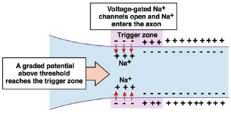
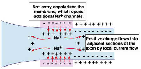
- Propagation of nerve impulses along the axon results from the diffusion of Na+ ions from the area that was just depolarized to the neighbouring area that is still polarized inside the axon
- When a part of the axon depolarizes, the localized are inside the axon becomes more positive as Na+ diffuses into the axon through voltage gated channels
- Outside the axon the concentration of Na+ is less in the depolarized region, so sodium diffuses from the polarized region towards the depolarized region
- The adjacent area inside the axon that is still polarized (more negative)
- The higher concentration of Na+ inside the depolarized region diffuses towards the polarized (more negative) region inside the axon
- These local currents causes the adjacent region to become more positively charged.
- When this happens, the membrane potential of the adjacent region becomes more positive from -70mv to -50mV (threshold potential)
- This results in a depolarization in the neighbouring region, as Na+ voltage-gated channels open and Na+ diffuses into the axon
6.5.U7 Synapses are junctions between neurons and between neurons and receptor or effector cells. [Only chemical synapses are required, not electrical, and they can simply be referred to as synapses.] (Oxford Biology Course Companion page 324)
- Define synapse, synaptic cleft and effector.
- State the role of neurotransmitters.
Synapses are junctions or structures between the pre-synaptic and post-synaptic membrane of two cells in the nervous system
- The junction can be between a neuron and an effector such as a muscle or a gland
- It can be between two different neurons. Many of these connections occur in the CNS (brain and spinal cord)
- A junction also exists between the sense receptor cells and the sensory neurons
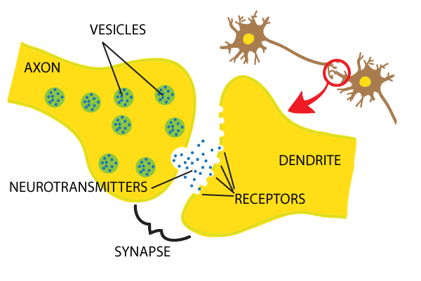
- Neurotransmitters are chemicals diffuse across a synapse from pre-synaptic membrane to post-synaptic membrane to send a signal to the next cell
6.5.U8 When presynaptic neurons are depolarized they release a neurotransmitter into the synapse.(Oxford Biology Course Companion page 325)
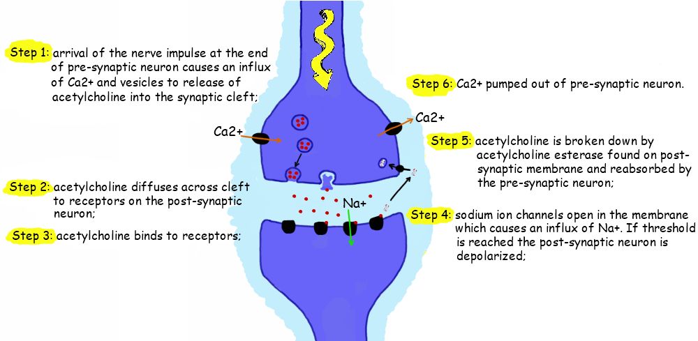
- Outline the mechanism of synaptic transmission, including the role of depolarization, calcium ions, diffusion, exocytosis, neurotransmitters, receptors, sodium ions, sodium channels, threshold potential and action potential.
Neurotransmitters are chemical messengers released from neurons and function to transmit signals across the synaptic cleft. Neurotransmitters are released in response to the depolarisation of the axon terminal of a presynaptic neuron
Neurotransmitters bind to receptors on post-synaptic cells and can either trigger (excitatory) or prevent (inhibitory) a response
Neurotransmitters bind to receptors on post-synaptic cells and can either trigger (excitatory) or prevent (inhibitory) a response
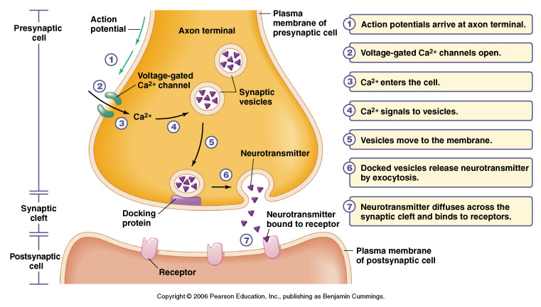
- As the nerve impulse reaches the axon terminal of the presynaptic neuron, thepositive charge from the depolarization causes voltage-gated channels permeable to Ca2+ to open.
- Ca2+ flows into the presynaptic neuron increasing the amount of Ca2+ in the presynaptic neuron.
- This Ca2+ causes vesicles containing neurotransmitters to bind to the membrane and release their neurotransmitters into the synaptic cleft (space between pre and post synaptic neuron).
- These neurotransmitters diffuse across the synaptic cleft and bind to receptor sites on the membrane of the post synaptic neuron.
- The binding of these neurotransmitters open ion channels allowing ions such as Na+ to diffuse into the post synaptic neuron.
- This influx of positive charge possibly leads to an action potential and adepolarization in the post synaptic neuron.
- The neurotransmitter is reabsorbed by the presynaptic neuron or broken down in the synapse by enzymes.
U 6.5.9 A nerve impulse is only initiated if the threshold potential is reached. (Oxford Biology Course Companion page 327)
- Outline the role of positive feedback and sodium ions in the reaching of threshold potential.
- Explain why some synaptic transmissions will not lead to an action potential in a postsynaptic cel
Action potential is only initiated if threshold potential is reached. Only at this potential does the voltage-gated sodium channels start to open, causing depolarization. Inward diffusion of sodium ions increases membrane potential causing more sodium channels to open (positive feedback effect). If the threshold potential is not reached, the post-synaptic membrane does not depolarize (sodium ions are pumped out and membrane returns to resting potential)
Typical post-synaptic neuron in the brain has synapses with many pre-synaptic neurons. This may be necessary for several of these to release neurotransmitter at same time for threshold potential to be reached and nerve impulse to be initiated in post-synaptic neuron (helps in decision making).
The threshold potential is the critical level to which a membrane potential must be reach in order to initiate an action potential
- Neurons fire or a nerve impulse is generated by an “all or nothing”
- When a stimulus occurs, some Na+ channels open causing the membrane potential to become more positive
- If enough Na+ diffuses into the neuron (-50mV to -70mV) and action potential is generated
- At a synapse, binding of a neurotransmitter at the post-synaptic membrane causes Na+ to diffuse into the neuron (if excitatory)
- This can cause a depolarization of the neuron if enough neurotransmitters are released
Application
6.5 A1 Secretion and reabsorption of acetylcholine by neurons at synapses. (Oxford Biology Course Companion page 326)
- Outline the secretion, action, reabsorption and formation of acetylcholine.
One example of a neurotransmitter used by both the central nervous system and peripheral nervous system is acetylcholine
- It is commonly released within the autonomic nervous system to promote parasympathetic responses (‘rest and digest’)
- It is largely used at the neuromuscular junction, meaning it is released by motor neurons and binds to receptors on muscles
- It is also used in the autonomic nervous system
- Acetylcholine is created in the presynaptic terminal by combining a water soluble nutrient called choline with an acetyl group
- Acetylcholine is secreted by the presynaptic membrane of a neuron
- The neurotransmitter diffuses across the synapse and binds to a receptor on the post synaptic membrane (causing an action potential if a threshold is reached)
- Once it has released from the receptor, an enzyme called acetylcholinesterase breaks down into choline and acetate
- Choline is reabsorbed back into the pre-synaptic neuron where it is combined with another acetyl group to form another acetylcholine neurotransmitter
6.5.A2 Blocking of synaptic transmission at cholinergic synapses in insects by binding of neonicotinoid pesticides to acetylcholine receptors. (Oxford Biology Course Companion page 326)
- Outline the mechanism of action of neonicotinoids use as insecticides.
- Define cholinergic synapse.
- Compare the proportion of cholinergic synapses in insects and humans.
- State why neonicotinoids insecticides are not highly toxic to humans.
The transmission of signals across a synapse can be slowed or blocked through a number of mechanisms, most of which prevent the neurotransmitter from binding to its receptor. Neonicotinoids are an example of this as molecules in the class are able to bind to acetylcholine receptors on post-synaptic cells in insects.
- Neonicotinoids bind to acetylcholine receptors in cholinergic synapses in the CNS of insects
- Acetylcholinesterase does not break down neonicotinoids therefore binding is irreversible
- Acetylcholine now can’t bind and neural transmission is stopped
- The insects go through paralysis and then death
- A benefit to this pesticide is that it is very effective in killing pests and it is not highly toxic to humans and mammals
- The problem is that it also effects beneficial insects such as honey bees. There is conflicting evidence if this is the case or not
- Many places such as the EU and Ontario, Canada has banned neonicotinoid pesticides
Skill
6.5.S1 Analysis of oscilloscope traces showing resting potentials and action potentials (Oxford Biology Course Companion page 324)
- Outline the use of oscilloscopes in measuring membrane potential.
- Annotate an oscilloscope trace to show the resting potential, action potential (depolarization and repolarization), threshold potential and refractory period.
Oscilloscopes can be used to measure the membrane potential of a neuron region over time by placing an electrode on each side of the membrane. As shown below, before an action potential occurs, the potential is at the resting value of -70mv. Then it spikes to +30 as the action potential passes and returns to the resting value after repolorization.
Key Terms:
|
peripheral
PNS dendrites cell body nodes of Ranvier propagating wave sodium channel presynaptic neonicotinoid refractory period exocytosis acetylcholinesterase |
nerves
neuron motor nerve impulse axon action potential propagation synapse acetylcholine calcium ions neonicotinoids |
active transport
sodium-potassium pump resting depolarisation repolarisation axon saltatory conduction synaptic cleft Schwann cells diffusion insecticides cholinergic synapse |
neurotransmitter
voltage-gated myelin synapse CNS arterioles vesicle ion channels depolarization threshold potential positive feedback local current |
impulse
effector terminal button synaptic terminal resting potential sodium ions potassium ions potassium channel receptors repolarization postsynaptic cell oscilloscopes |
PowerPoint and Notes on Topic 6.5 by Chris Payne
Your browser does not support viewing this document. Click here to download the document.
Your browser does not support viewing this document. Click here to download the document.
Correct use of terminology is a key skill in Biology. It is essential to use key terms correctly when communicating your understanding, particularly in assessments. Use the quizlet flashcards or other tools such as learn, scatter, space race, speller and test to help you master the vocabulary.
External Links:
Control, regulation and feedback
Link to Neurobiology and Behaviour: E4 – Neurotransmitters and Synapses
Loads of useful resources from Neuroscience for Kids
Reflex arc animation from msjensen.
Nerve impulses: resting potential and action potential
Cool tutorial from the Harvey Project
McGraw Hill nerve impulse animation
Nice and simple from mrothery
PhET Lab simulation: Neurons (allow Java to run)
Another good one from Alberta Psychology
Synaptic Transmission
Great animation from McGraw Hill
Good tutorial from Harvard Outreach
Another WHS Freeman tutorial (lifewire)
In the News:
Which One Will Make You Fat
New Insights Into Brain Neuronal Networks from Science Daily
Neuroscience For Kids
Insect gene expression responds to diet from BrightSurf
Video Clips:
Hank begins a series of videos on organ systems with a look at the nervous system and all of the things that it is responsible for in the body.
Hank goes through a discussion of the nervous system
Paul Andersen begins this podcast with a discussion of brain lateralization and gives a brief demonstration of tests that were performed on split-brain individuals. He then discusses the major parts of a neuron and explains how action potentials are generated using voltage-gated ion channels. He explains how neurotransmitters transmit messages across a synapse and how these messages can be either inhibitory or excitatory.
Good explanation of action potential
In my 2-Minute Neuroscience videos I explain neuroscience topics in 2 minutes or less. In this video, I discuss the action potential. The term "action potential" refers to the electrical signaling that occurs within neurons. This electrical signaling leads the release of neurotransmitters, and therefore is important to the chemical communication that occurs between neurons. Thus, understanding the action potential is important to understanding how neurons communicate.
All thought and action result from your nervous system: the brain, spinal cord and nerves. This system is more sophisticated than any computer
In my 2-Minute Neuroscience videos I explain neuroscience topics in 2 minutes or less. In this video, I discuss synaptic transmission. I describe the synapse, synaptic cleft, release of neurotransmitter and its interaction with receptors, and the ways neurotransmitter is cleared from the synaptic cleft.
The Centre for Ecology & Hydrology (CEH) is undertaking research to quantify the impact on honeybees of two commercial neonicotinoids seed treatments in commercially grown crops of oilseed rape
Making memories
Dr. Chiaravalloti discusses the learning process and techniques that have been shown to improve learning and memory in healthy persons. She walks through the evidence demonstrating these techniques to additionally be helpful to persons with Multiple Sclerosis and Traumatic Brain Injury.
